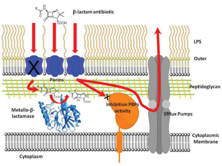
β-Lactam antibiotics are the most widely used chemotherapeutic agents for the treatment of bacterial infections. The main mechanism of resistance to these drugs is the synthesis of β-lactamases, enzymes that hydrolyze and inhibit the action of these antibiotics. Metallo-β-lactamases (MβLs) constitute the most recent generation of these enzymes. MβLs have a broad spectrum of action, inactivating all β-lactam antibiotics, including carbapenems (the last resort used in clinical practice).
The dissemination of genes encoding these enzymes in opportunistic and pathogenic organisms is now global. This situation is exacerbated by the structural diversity of the different MβLs, which makes it difficult to design an efficient inhibitor for these enzymes. The NDM-1 enzyme (http://www.bbc.co.uk/news/health-10925411), in particular, is rapidly spreading worldwide, and there are no clinically used inhibitors for these enzymes, posing a threat to global health.
Our goal is to elucidate the structure-function relationship of these enzymes through biochemical, structural, mechanistic, and evolutionary studies, with the ultimate goal of designing a clinically applicable inhibitor. To date, we have succeeded in: (1) proposing a common catalytic mechanism for MβLs by characterizing a reaction intermediate, (2) identifying their functional in vivo species, and (3) exploring their potential evolutionary mechanisms. We are currently using this prior knowledge to design and generate MβL inhibitors, under the hypothesis that, despite their structural diversity, MβLs act using the same catalytic mechanism.
Our group conducts an interdisciplinary study, using techniques from molecular biology, biochemistry, structural biology, and modern enzymology. Mechanistic studies are performed using rapid mixing techniques and various spectroscopies to follow changes in the active site during catalytic turnover on the millisecond timescale, with the goal of identifying reaction intermediates.
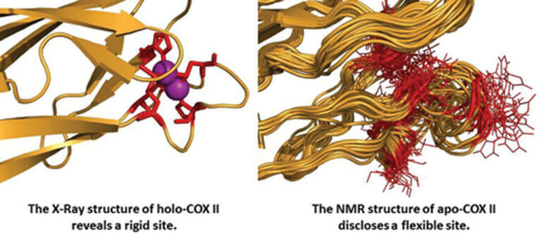
We use crystallography and/or Nuclear Magnetic Resonance (NMR) to characterize the binding mode of potential inhibitors to these enzymes. We also use in vitro directed evolution strategies as a means to predict the evolution of these enzymes in hospital environments and to understand how mutations are fixed in the evolutionary process.